Multimodal electron microscopy enables comprehensive insights into organic solar cells
How do organic solar cells work on the inside? The answer lies in structures far too small to see—and difficult to access even with advanced techniques. So far, researchers have relied mainly on X-ray methods to understand how molecules are arranged within these materials and how this order can be optimized for high efficiency. While powerful, X-rays provide only a spatially averaged picture. Electrons, in contrast, offer a local view at the nanoscale, revealing both structure and chemical composition. A complete analysis has therefore required combining both approaches. In a new study, researchers at Friedrich-Alexander-Universität (FAU) Erlangen-Nürnberg, together with partners from Forschungszentrum Jülich and DESY in Hamburg, now show that electrons alone can achieve both. The work has been published in the renowned journal Nature Communications.*
Using three-dimensional electron diffraction (3D ED), they demonstrate that electrons can also provide the averaged structural information previously accessible only with X-rays. “For the first time, this allows a comprehensive structural characterization of organic solar cells within a single instrument—a transmission electron microscope,” says Prof. Erdmann Spiecker, head of the Institute of Micro- and Nanostructure Research and the Center for Nanoanalysis and Electron Microscopy (CENEM) at FAU, who led the project. “3D ED was the missing piece we needed to complete the electron microscopy toolbox.”
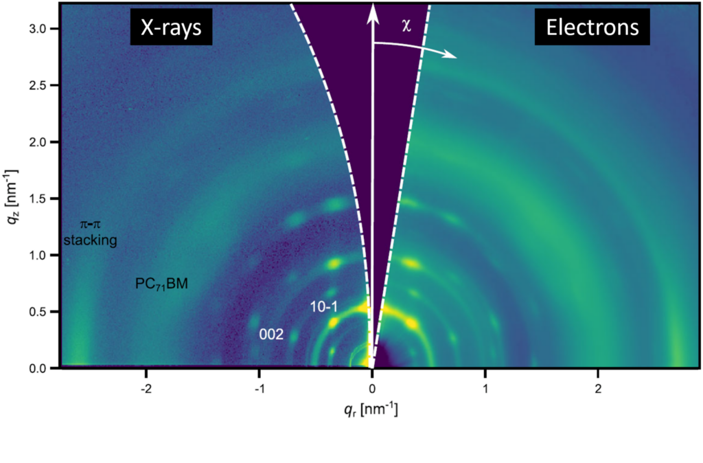
At the heart of the work is a direct comparison of electron and X-ray data from identical samples. “It is striking how well the data agree, despite the fundamentally different nature of electrons and X-rays,” says Irene Kraus, who carried out the 3D ED experiments as part of her doctoral research. The measurement geometries differ markedly: X-rays probe the sample at shallow angles in reflection, while electrons pass through it. “In 3D ED, we tilt the sample step by step, allowing us to reconstruct the average molecular order in three dimensions—much like tomography.”
A key challenge is radiation damage. Organic solar cells are extremely beam-sensitive, and even low doses can disrupt their delicate molecular order. This is particularly critical for electrons, which interact strongly with matter and deposit energy locally. “At first glance, electrons may seem unsuitable for such materials,” says Dr. Mingjian Wu, senior scientist at CENEM, who co-supervised the project. “But with careful dose control, we can extract structural information before damage occurs.” By optimizing the electron dose and developing tailored acquisition strategies, the team was able to reliably probe even highly sensitive nanocrystalline structures—capturing detailed structural information while preserving the material.
Importantly, the results do not diminish the role of X-ray techniques. Rather, the methods are complementary. X-rays require minimal sample preparation and are particularly well suited for in situ studies of structural evolution during processing. Electron microscopy, in contrast, now uniquely combines averaged structural information with local imaging, diffraction imaging and chemical analysis in a single instrument. “This is what we call multimodal microscopy,” explains Spiecker. “It allows us to directly link different types of information—from molecular texture to local order and composition—within one experiment.” Multimodal and correlative microscopy of functional materials are a major research focus at CENEM. This approach lies at the heart of the recently established DFG-funded Research Training Group RTG 3103 “Correlative Materials Microscopy: From nanostructured functional films to hierarchical functional materials (CorMic)”, a doctoral program comprising 13 PhD projects. The insights gained into solar cell materials are also central to the DFG-funded Collaborative Research Center CRC 1719 “Next-generation printed semiconductors: Atomic-level engineering via molecular surface chemistry (ChemPrint)”. Both initiatives are hosted by FAU Erlangen-Nürnberg.